Abstract
Neurofeedback (NF) is a type of biofeedback in which an individual’s brain activity is measured and presented to them to support self-regulation of ongoing brain oscillations and achieve specific behavioral and neurophysiological outcomes. NF training induces changes in neurophysiological circuits that are associated with behavioral changes. Recent evidence suggests that the NF technique can be used to train electrical brain activity and facilitate learning among children with learning disorders. Toward this aim, this review first presents a generalized model for NF systems, and then studies involving NF training for children with disorders such as dyslexia, attention-deficit/hyperactivity disorder (ADHD), and other specific learning disorders such as dyscalculia and dysgraphia are reviewed. The discussion elaborates on the potential for translational applications of NF in educational and learning settings with details. This review also addresses some issues concerning the role of NF in education, and it concludes with some solutions and future directions. In order to provide the best learning environment for children with ADHD and other learning disorders, it is critical to better understand the role of NF in educational settings. The review provides the potential challenges of the current systems to aid in highlighting the issues undermining the efficacy of current systems and identifying solutions to address them. The review focuses on the use of NF technology in education for the development of adaptive teaching methods and the best learning environment for children with learning disabilities.
Keywords:
1. Introduction
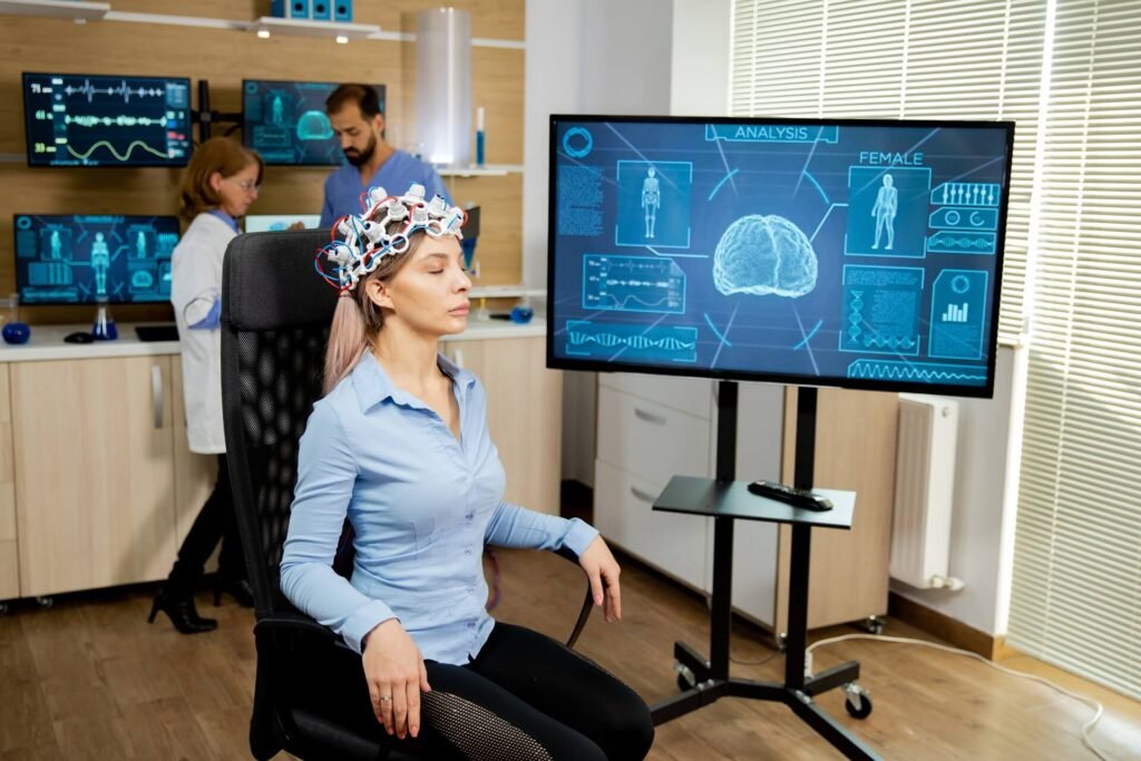
Neurofeedback (NF) is a type of biofeedback, the most common and traditional form of which is the electroencephalogram (EEG) NF (EEG-NF). EEG-NF displays EEG waves that are measured via electrodes placed on the scalp. The brain is constantly active, whether one is awake or asleep and whether in the presence or absence of distinct stimuli. EEG waves reflect the brain’s current functional state and can be characterized and separated into delta, theta, alpha, sensorimotor rhythm (SMR), beta, and gamma frequency bands. Each band is a unique indicator of brain activity. For example, brain activity during a complex cognitive task varies from that at rest, and the strengths of the EEG bands vary correspondingly. EEG-NF training captures characteristic EEG bands in real time and also measures the changes in EEG activity that are critical for and promote changes in a targeted cognitive function. In recent years there has been a significant increase in clinical applications of the technique for several neuropsychiatric conditions, including attention-deficit/hyperactivity disorder (ADHD), epilepsy, migraine, and headaches [1,2,3,4,5].
NF methods are usually based on EEG, functional magnetic resonance imaging (fMRI), or near-infrared spectroscopy (NIRS). Hitherto, EEG has been the primary method used in NF, mainly because of its low cost, ease of use, and non-invasiveness [6]. Technological advances have now made it possible to combine methods, as in the simultaneous EEG-fMRI method in which the subject wears an EEG electrode cap while inside an MRI scanner. In this advanced method, EEG and fMRI recordings are performed simultaneously [7]. Advances in fMRI-NF have demonstrated promising results with techniques such as decoded NF (DecNef) and functional connectivity NF (FCNef) [8,9,10,11]. In the current report, we restrict our focus to EEG-NF.
EEG comprises a mixture of multiple waves of various frequencies. These frequency bands are correlated with various degrees of neuronal synchronizations [12] and are also linked to a variety of cognitive operations (Harmony, 2013). For example, delta waves (0.5–4 Hz) are associated with inhibition of the sensory afferents [13,14]; theta waves (4–8 Hz) are typical of nervousness [15,16]; an attentive and relaxed state of mind is characterized by alpha waves (8–12 Hz) [17,18,19]; alertness is characterized by beta waves (16–30 Hz) [20]; and problem-solving or higher cognitive functions are associated with gamma waves (30–100 Hz) [21,22]. In terms of resting-state EEG frequency bands, waking state eyes-open activity is correlated with beta waves, whereas stage I non-rapid eye movement (non-REM) sleep is characterized by theta waves. Stage II non-REM sleep is characterized by spindles in the 10–15 Hz frequency range, whereas stage III and IV non-REM sleep are characterized by slow delta waves. Rapid eye movement (REM) sleep, after this stage, is characterized by high frequency and low amplitude waves, which is similar to the brain activity when individuals are awake.
Some characteristics of the EEG components and their frequency ranges are presented in Table 1. EEG-NF protocols usually depend on the power of various EEG frequencies such as the delta, theta, alpha, and beta bands or the alpha/theta or beta/theta ratios [23].
Table 1. Characteristics of EEG frequency bands.

NF systems consist of five basic components, as illustrated in Figure 1. The first is the data acquisition component, which is used to acquire brain signals. Neural responses can be recorded using EEG, electrocorticography (ECoG), intracranial EEG, or magnetoencephalography (MEG). The advantage of using these methods for NF is their high temporal resolution, which implies that brain activities are continuously and directly reflected within milliseconds. High spatial resolution fMRI and near-infrared spectroscopy (NIRS) are also being increasingly used for data acquisition. The second component is online data processing, which involves the detection and removal of artifacts from the signals provided by the data acquisition component. Typical artifacts from EEG/ECoG/MEG include muscle or eye movements, and 50/60 Hz power line interference. Further to this, artifacts associated with EEG at the scalp are a mix of a spatial smearing effect from the skull and other tissues and multiple sources of brain activity [24]. Placing electrodes close to each other nearby reduces the number of artifacts and results in a better signal from the scalp. The blind source separation (BSS) method is typically used to separate artifacts from signals such as the EEG [25,26,27]. This method detects signal sources that are important for understanding brain activity [28]. In fMRI-based NF, participants attempt to control their brain activity using real-time feedback from a particular region or network [29,30]. The most common artifacts in the NIRS/fMRI signal are head motion, body motion, respiration, and heartbeat. Artifacts caused by inhomogeneity in the magnetic field are also common in fMRI-NF. Similar to the case of EEG, good signal processing methods such as BSS can separate these artifacts from fMRI signals [31].


